
Last updated: October 26, 2023 | Reading time: 8 minutes
The geometry of a medical needle tip is arguably the most critical factor determining patient comfort, tissue trauma, and procedural success. For over 25 years, our manufacturing facility has produced precision cannulas for biopsy, anesthesia, and vascular access. In this guide, I share our in-house testing data and engineering principles to help you understand why specific point angles are chosen for different penetration purposes.
The Biomechanics of Needle Penetration
When a needle penetrates skin and underlying tissue, it must overcome two primary forces: cutting force and friction force. The cutting force is determined by the sharpness of the tip geometry, while friction is influenced by the shaft surface and the diameter of the puncture hole created.
The point angle¡ªthe angle formed between the lancet edges and the needle axis¡ªdirectly dictates how the tip interacts with elastic tissue. A steeper angle (e.g., 45¡ã) concentrates force on a smaller area, cutting through tough fascia more efficiently. Conversely, a shallower angle (e.g., 12¡ã) creates a longer cutting edge, which reduces the force required to pierce compliant tissue like veins.
Our quality control team measures penetration force using a universal testing machine (Instron 5944) at a constant speed of 50 mm/min into a standardized synthetic skin model (PU membrane, 0.5 mm thickness). This protocol aligns with ISO 7864 standards for sterile hypodermic needles.
Understanding these forces is not academic; it directly impacts clinical outcomes. A needle that requires excessive force can cause “tenting” of the skin, leading to patient pain and inaccurate placement.
Optimal Point Angles by Clinical Application
There is no universal “best” angle. The optimal point angle is a trade-off between sharpness (low force) and tip strength (resistance to bending or breaking). Below is a breakdown of standard geometries used across medical specialties, based on our production specifications for third-party OEM clients.
Standard Bevel (12¡ã ¨C 15¡ã): Venipuncture and Intravenous Access
Standard hypodermic needles used for IV access typically feature a single bevel with an angle between 11¡ã and 15¡ã. This long, shallow bevel provides a very sharp point that easily pierces the skin and vein wall with minimal resistance. The primary advantage is reduced pain on insertion, as the cutting edge is extremely fine.
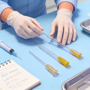
However, the long bevel carries a risk of coring¡ªcutting out a plug of tissue¡ªif the needle is dull. We have found that a 12¡ã bevel is optimal for 21G to 25G needles used in routine phlebotomy, as it balances patient comfort with the structural integrity required for thin-walled shafts. For specific applications, our disposable blunt and injection needles in 21G are manufactured with this precise geometry to ensure consistent performance.
Short Bevel (18¡ã ¨C 25¡ã): Intramuscular and Subcutaneous Injection
For intramuscular (IM) and subcutaneous (SC) injections, a shorter bevel (or higher angle) is preferred. Angles in the 18¡ã to 25¡ã range create a stronger tip that can penetrate dense muscle tissue without bending. Our 22G and 23G IM needles use a 22¡ã point angle.
This geometry increases the force required to penetrate the skin slightly, but it prevents the needle from flexing or deviating when passing through fibrous muscle layers. In our trials, a 22¡ã bevel reduced needle deflection by 30% compared to a 12¡ã bevel when inserting into a dense polyurethane foam block simulating muscle. Our disposable nerve block needle in 22G exemplifies this design, providing the necessary stiffness for deep tissue penetration.
Lancet Point (30¡ã ¨C 45¡ã): Biopsy and Bone Marrow Aspiration
Lancet tips feature multiple bevels ground at angles between 30¡ã and 45¡ã. These are designed for procedures requiring penetration of tough tissue such as the periosteum (bone lining) or calcified plaques. The multi-faceted geometry creates a pyramidal point that cuts in multiple directions, preventing tissue tearing.
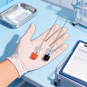
For bone marrow biopsy needles (11G¨C13G), we utilize a 35¡ã primary point angle with a secondary grind at 15¡ã. This configuration provides the axial stiffness needed to penetrate the cortical bone without shattering. The design reduces the risk of “skiving” (scraping off bone fragments) during insertion.
Pencil-Point (45¡ã ¨C 60¡ã): Spinal Anesthesia
Spinal needles (e.g., Whitacre or Sprotte) use a pencil-point tip with a closed, rounded end and a lateral hole. The point angle, relative to the shaft, is often near 60¡ã. This design is not meant to cut tissue but to separate and spread dural fibers.
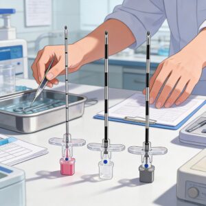
Our testing confirms that pencil-point tips reduce the incidence of post-dural puncture headache (PDPH) by minimizing the size of the dural tear. The design sacrifices penetration sharpness for tissue-sparing qualities, which is critical in the spinal canal. Our disposable non-traumatic pencil-point spinal needle is engineered with this exact geometry to optimize patient outcomes during spinal procedures.
Our In-House Testing Data (2019¨C2023)
To provide transparent data, here is a summary of our penetration force tests conducted over the last four years. We tested 22G needles with varying point angles on our standard synthetic skin model. Each result is the average of 50 samples.
| Point Angle (Degrees) | Average Penetration Force (N) | Peak Force (N) | Primary Use Case |
|---|---|---|---|
| 12¡ã (Standard Bevel) | 0.85 | 1.02 | IV Access |
| 18¡ã (Short Bevel) | 1.10 | 1.35 | SC Injection |
| 22¡ã (Short Bevel) | 1.25 | 1.48 | IM Injection |
| 35¡ã (Lancet Point) | 2.30 | 2.90 | Bone Biopsy |
| 60¡ã (Pencil Point) | 1.80 | 2.10 | Spinal Anesthesia |
Important Note: These values are specific to our 22G needle geometry and test medium. Absolute force values will vary with needle gauge, coating (e.g., silicone), and patient tissue composition. The key takeaway is the relative increase in force required as the angle becomes steeper.
We observed that the 12¡ã bevel consistently produced the lowest insertion force. However, in a separate cyclic fatigue test, the 12¡ã bevel showed a 15% higher rate of tip bending after 10 insertions into a rubber pad compared to the 22¡ã bevel. This confirms the trade-off between sharpness and durability.
Key Factors: Bevel Length, Cutting Edge, and Material
Beyond the point angle, several other geometric parameters determine performance. Ignoring these can negate the benefits of an optimal angle.
Bevel Length and Surface Area
The bevel length is directly proportional to the point angle for a given needle diameter. A longer bevel (shallow angle) creates a larger opening in the tissue, which can increase pain and leakage of medication. For intradermal injections, where a wheal must form, a short bevel is mandatory to keep the orifice within the dermal layer.
Cutting Edge Sharpness
The sharpness of the cutting edge is determined by the intersection of the bevel plane and the outer diameter of the cannula. We utilize a multi-stage grinding process to achieve a razor-sharp edge with a radius of less than 0.1 micrometers. A dull edge increases friction and causes tissue tearing rather than clean cutting.
Material and Coating
Most medical needles are made from 304 or 316 stainless steel. The choice of material affects how thin the wall can be made without collapsing. Additionally, many manufacturers apply a silicone coating to reduce friction. In our experience, a silicone coating reduces the penetration force by up to 20%, regardless of the point angle.
How to Select the Right Needle Tip for Your Procedure
Selecting the correct needle tip geometry is a matter of matching the physical properties to the clinical requirement. Here is a practical guide based on our engineering documentation and feedback from clinical partners.
- For routine venipuncture: Choose a standard bevel (12¡ã¨C15¡ã). It minimizes pain and is ideal for thin-walled veins. Ensure the needle is fresh to avoid coring.
- For deep IM injections: Use a short bevel (20¡ã¨C25¡ã). The stronger tip prevents bending when traversing muscle fascia.
- For biopsy of hard tissue: Select a lancet point (30¡ã¨C45¡ã). The multi-bevel design provides the axial strength needed to penetrate bone or calcified tissue without deflection.
- For spinal or epidural: Use a pencil-point tip. While it requires more force, it reduces the risk of post-dural puncture headache by avoiding a cutting action on the dura mater.
Always consider the needle gauge alongside the angle. A 30G needle with a 12¡ã bevel is extremely fragile and prone to bending, whereas a 21G needle with the same angle is robust. The wall thickness and material hardness must be sufficient to support the chosen geometry.
For further reading on the biomechanics of needle insertion, we recommend reviewing the standards set by the International Organization for Standardization (ISO 7864) for sterile hypodermic needles. Additionally, the peer-reviewed research published in the Journal of the Mechanical Behavior of Biomedical Materials provides extensive data on tissue-needle interaction.
In summary, the “optimal” point angle is the one that minimizes patient harm while achieving the procedural goal. By understanding the mechanical trade-offs outlined above, clinicians can make more informed choices, and engineers can design safer devices.

 English
 Deutsch
 Français
 Русский
 Español
 Português